Striking New Paths with Traditional Ultrasound
How the Combination of Traditional Ultrasound and MotionCapturing can improve Clinical 3D Gait Analysis
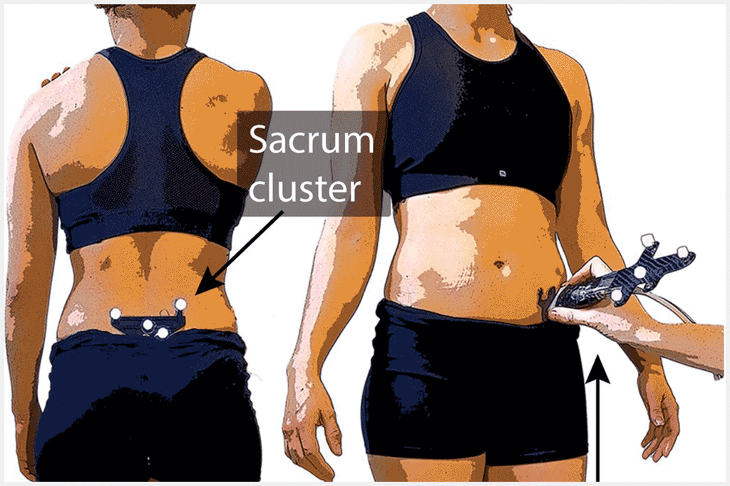
This study combines a traditional ultrasound device with a MotionCapture system to be able to “see” and quantify anatomical structures in 3D, based on two-dimensional ultrasound images.
The team of Brian Horsak applies this experimental method in an attempt to make clinical 3D gait analysis for children and adolescents suffering from obesity even more precise. For example, the approach allows to determine the 3D position of the femoral head, which would normally require an X-ray examination.
Project HIPstar
The publication was developed within the framework of the FWF-promoted project HIPstar, in which staff members of the St. Pölten UAS-based Institute of Health Sciences and Center for Digital Health Innovation collaborate with numerous renowned universities and partner clinics including
• the Orthopaedic Hospital Vienna-Speising
• the children’s hospital of the Medical University of Vienna
• the Karl Landsteiner University of Health Sciences
• the University of Vienna
• and KU Leuven in Belgium
Read the publication here (website of the publishing company Nature).